HELPING HAND TO LABORATORY
by Ekaterina ZININA, Vlada KAPUSTINA
Scientific innovations help to keep the equipment up and running – the one that may win several months in cancer detection outgoing any other diagnostic techniques.
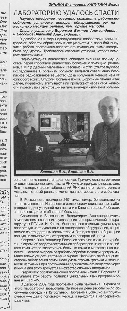
Helping hand was offered by Victor VORONKOV and Vladimir BESSONOV
It was in December 2007 when Nuclear Medicine Study Department in Kaliningrad Regional Hospital became looking for specialists to help them: the firmware of their gamma camera was at risk of failure. It was necessary to save the equipment that was saving lives of people.
Nuclear medicine technologies show great advantages in diagnostics over x-ray, nuclear magnetic-resonance (NMR) and ultra sound examination methods. A patient takes a special radioactive agent, which is absolutely harmless (radiation doze is less than the exposure during photofluorography). As various tumors, diseased kidneys, cirrhotic livers and other abnormalities have different capacity of absorption and infiltration of liquids, registration of the radiation of such affected organs by gamma camera allows detecting and measuring them. Nuclear Medicine Imaging enables to detect problems at early stage which is not always possible with X-raying. For certain diseases Nuclear Medicine Study is the only diagnostic technique that can be used.
Overall there are about 240 gamma cameras in Russia, the major part of those being heavily worn-out. This was also the case with Nuclear Medicine Study Laboratory in Kaliningrad Regional Hospital, which is in fact the only Nuclear Study Institution in the region.
Jointly with Vladimir Bessonov, Deputy Head of Information Infrastructure Department at the Immanuel Kant State University of Russia it was decided to replace the unique hardware of the aggregate by standard computer compatible equipment. This idea became the key that made the Laboratory no more dependent on any software and hardware.
By April 2009 V. Bessonov completed his share of work. To great fanfare of the Lab staff they could see the diseased kidneys and metastases of their patients at the monitor which was connected to their off-the-shelf computer. It then became necessary to develop data processing software. Because it is not enough to see the image displayed at the screen. For example to assess how seriously a kidney is affected one should be able to make a radiation intensity graph that shows how the radiant agent passes through this kidney. This requires the development of many complex algorithms.
The development of data processing program was undertaken by V. Voronkov. This came along with the development of the unique FORT-VV programming language.
The software development was completed by December 2009. In February the Laboratory has come back to life. During its first working day 12 patients have been examined. The program has now been used for more than two months and it is in the process of ongoing development.
On the photo: V. Bessonov, V. Voronokov
|